PBSC = Peripheral Blood Stem Cell
91 case patients x 10 mL whole blood [whole blood donor Jennifer Joy Brenley Nyhof] x 2 doses per protocol patient all allocated and administered with regards to clinical trials commencing August 2024 and ongoing currently, and finished in follow-up forward-looking September – October 2025, all with respect to the following:
Nyhof, Jennifer Joy Brenley. Self-Published. (2026) Evidence for the cure of human immunodeficiency virus (HIV) infection through autologous whole blood cell transfusion via a mechanism of donor-derived and D-DOPA catalyzed human immunoglobulin IgD-IgA1 dimerization and subsequent virus-specific immune complex formation in the recipient host with acknowledgement of donor-provided complement fixation and membrane attack complex (MAC) formation in-situ, and with special emphasis placed on C2 in this context.
Nyhof, Jennifer Joy Brenley. Self-Published. (2026) Evidence for the cure of acute lymphoblastic leukemia (ALL) through autologous whole blood cell transfusion via a mechanism of donor-derived and D-DOPA catalyzed human immunoglobulin IgD-IgA1 and human immunoglobulin IgD-IgA4 dimerization and subsequent neoplastic cell-specific immune complex formation in the recipient host with acknowledgement of donor-provided complement fixation and membrane attack complex (MAC) formation in-situ, and with special emphasis placed on C2 and C3 in this context.
Nyhof, Jennifer Joy Brenley. Self-Published. (2026) Evidence for the cure of glioblastoma (GBI) through autologous whole blood cell transfusion via a mechanism of donor-derived and D-DOPA catalyzed human immunoglobulin IgD-IgA1 dimerization and subsequent neoplastic cell-specific immune complex formation in the recipient host with acknowledgement of donor-provided complement fixation and membrane attack complex (MAC) formation in-situ, and with special emphasis placed on C2 in this context.
Nyhof, Jennifer Joy Brenley. Self-Published. (2026) Evidence for the cure of retinoblastoma (RBB) through autologous whole blood cell transfusion via a mechanism of donor-derived and D-DOPA catalyzed human immunoglobulin IgD-IgA2 and human immunoglobulin IgG4-IgG2 dimerization and subsequent neoplastic cell-specific immune complex formation in the recipient host with acknowledgement of donor-provided complement fixation and membrane attack complex (MAC) formation in-situ, and with special emphasis placed on C2 and C3 in this context.
Nyhof, Jennifer Joy Brenley. Self-Published. (2026) Evidence for the cure of mammary gland adenocarcinoma (MGAC) through autologous whole blood cell transfusion via a mechanism of donor-derived and D-DOPA catalyzed human immunoglobulin IgD-IgA2 and human immunoglobulin IgG4-IgG2 dimerization and subsequent neoplastic cell-specific immune complex formation in the recipient host with acknowledgement of donor-provided complement fixation and membrane attack complex (MAC) formation in-situ, and with special emphasis placed on C2 and C3 in this context.
Nyhof, Jennifer Joy Brenley. Self-Published. (2026) D-DOPA, the more useful and biologically active enantiomer of DOPA, as an enzyme catalyst and neurotransmitter for the primary, secondary, and tertiary prevention of Parkinson’s Disease.
Nyhof, Jennifer Joy Brenley. Self-Published. (2026) D-DOPA, the more useful and biologically active enantiomer of DOPA, as an enzyme catalyst and neurotransmitter for the primary, secondary, and tertiary prevention of thromboembolic disease [cerebrovascular infarct (C.V.I.), transient ischemic attack (T.I.A.), pulmonary thromboembolism (PTE), deep vein thrombosis (DVT), disseminated intravascular coagulation (D.I.C.)]
Introduction
- Preamble
- D-penicillAMINE, penicillAMINE D-enantiomer [external link, Wikipedia nice work]
- 3,4-dihydroxy-d-phenylalanine [D-DOPA] c.f. 3,4-dihydroxy-l-phenylalanine [L-DOPA] [stereochemistry]
- D-DOPA physical, chemical, and biological properties and characteristics [internal link]
- (F-D-DOPA PET/CT Scan Fluoro-DOPA d-enantiomer specific nuclear medicine nigrostriatal pathophysiology positron emission tomography computed tomography)
- L-DOPA is the WRONG ANSWER and L-DOPA is a toxic fungal metabolite [internal link]
- D-DOPA physical, chemical, and biological properties and characteristics [internal link]
- Tyrosine Hydroxylase 2SXN 191290 enzyme D-specific [enantiomer chirality][non-superimposable mirror images about a chiral carbon][polarized light]
- Features of the tyrosine hydroxylase enzyme [internal link]
- Segawa Syndome, autosomal recessive inheritence [gene-phenotype relationship]; cytogenetic location: gene/locus on chromosome 11 [11p15.5]
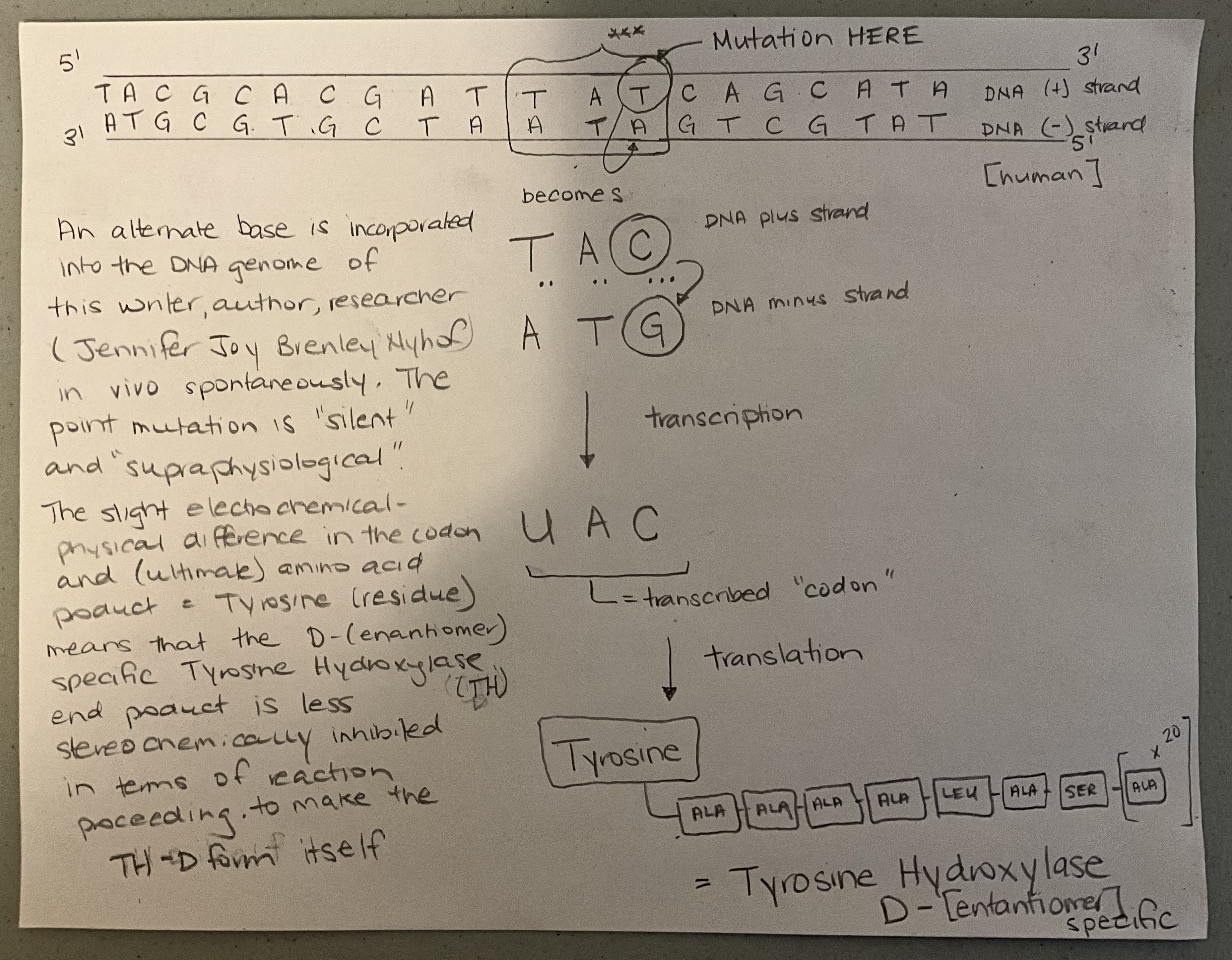
- TAC – ATG —> UAC “silent” (supraphysiologic) in-vivo, spontaneous, point mutation in the DNA genome of the writer, author, researcher (human female, Jennifer Joy Brenley Nyhof) leads to a product of translation that is a less stereochemically inhibited Tyrosine residue and, amongst other things, formation of d-enantiomer specific Tyrosine Hydroxylase is more favorable, relatively speaking, compared to all other humans Homo sapiens species. See diagram.
- Short Tandem Repeats STR’s are abnormal findings in context of “genome mapping” all species considered and this writer author, Jennifer Joy Brenley Nyhof, does not have STR’s in her genetic profile (genome) and the STR regions that are present in the genome DNA of other humans (not me) represent “missed opportunities”, as they are the “coding regions” that otherewise would result in D-DOPA production in terms of the “final outcome” proteomic scenario [with reference to the FBI Federal Bureau of Investigation CODIS Combined DNA Index Database]
- ΔG Gibbs Free Energy (zinc Zn reaction center) = -4312.9967121
- ΔG Gibbs Free Energy (zinc Zn reaction center)D-DOPA catalyst = -21.67912587
- [immunoglobulin dimerization and getting the job done]
- antiseroconversion
- nucleic acid test gold standard PCR polymerase chain reaction
- human immunodeficiency virus (HIV) induced cytopathology (Lang et al. 2011; Lu et al. 2021) happens (HIV infected cells are not normal in how they look or behave) and this occurs in the case of HIV+ patients / recipients who are in need of the whole blood product that contains D-DOPA and dimerized immunoglobulins (antibodies) IgD-IgA1 and healthy receptor-specific T-cells and Natural Killer Cells (NKC’s) and primed and ready macrophages from the healthy whole blood donor (the healthy whole blood donor is Jennifer Joy Brenley Nyhof [internal link] and the cells and antibodies in the energized whole blood hyperimmune product that people get from me (JJBN blood donor) “recognize” the cytopathological “diseased-looking” and “diseased-acting” infected cells in the people receiving the “transfusion” and targets the infected cells for destruction and then the viral-infected cells are killed like destroyed
- Viral replication within the cells of HIV+ patients happens in 4 minutes after virus uncoating and 18-24 new viruses are made in the 97.1281 seconds later after the 4 minute time halt
- in a newly infected patient that “catches” the HIV “virus” – their immune system can’t do any better than killing a viral infected cell 19 minutes later and after 81-97 copies (new viruses) have been made
- hyperimmune D-DOPA “energized” (enzyme catalysis) whole blood from HEALTHY whole blood (non-HIV infected HIV negative Jennifer Joy Brenley Nyhof) donor TRANSFUSED into the HIV+ “sick” patient per protocol (see later) kills HIV infected cells in 8 seconds after viral uncoating (BEFORE viral replication can take place)
- EX-VIVO / IN-VIVO [see T1/2 half life numbers written out later in this same research paper list segment that you are reading right now]
- whole blood from “any” healthy donor (not hyperimmune) who is of the AB+ blood (control donor group AB+) transfused into an HIV positive person is ineffective at producing antiseroconversion and cell death of a viral infected cell takes 9 minutes (and recall that viral replication takes 4 minutes)
- whole blood from “any” healthy donor (not hyperimmune) who is of the O- blood (control donor group O-) transfused into an HIV positive person is ineffective at producing antiseroconversion and cell death of a viral infected cell takes 10 minutes (and recall that viral replication takes 4 minutes)
- in a person who is HIV+ and in a situation that has progressed to AIDS acquired immunodeficiency syndrome, the (non-functioning) immune system takes 91 minutes to kill a virus infected cell
- A “kill time” of LESS than 3 minutes and 39 seconds is needed and ride ‘em cowboy ‘cause i get ‘em at 8s
- the “trick” is knowing and being able to distinguish self from non-self PRECISELY at the cellular level (even after the blood has left my body and is flowing through the veins and arteries of the recipient patient) and this is made possible by T-cells and natural killer cells and macrophages of the monocyte cell line and supraphysiological blood (serum) concentrations of D-DOPA and more things than this yes and B-cells some too [Rackaityte E and Halkias J (2020)]
- the half life of D-DOPA like the T1/2 in whole blood at (supra)physiological (body) temperature and pH conditions is 9.12 days
- the half life of IgD-IgA1 like the T1/2 in whole blood at (supra)physiological (body) temperature and pH conditions is 14.71 days
- IgD-Fc: Cδ3 domain [conformational change] (Davies et al. 2025)
- monomeric IgG4 (Oganesyan et al. 2016, Shan et al. 2016)
- CD38 cluster of differentiation 38 cell surface marker antigen
- Human CD (Cluster of Differentiation) Antigen Cell Surface Marker Information in Table and Chart format [The Immunology Link (2020)]
- CD1 – CD100 [external link]
- CD101 – CD200 [external link]
- CD201 – CD300 [external link]
- CD301 – CD339 [external link]
- Human CD (Cluster of Differentiation) Antigen Cell Surface Marker Information in Table and Chart format [The Immunology Link (2020)]
- C808 is a cell surface feature of life and it is expressed in a pre-mitotic spindle formation of fashion. It is denoted by a C and not a CD for obvious reasons. If the cell goes on to live and differentiate, this cell surface marker evolves into CD1b and it remains as an expression of “self” that is present on all cells of the human body (including red blood cells) and the CD1b cell surface marker induces ongoing “tolerance” with regards to the behavior and function of healthy immune system cellular and non-cellular components. A misshapen CD1b is the first sign symptom and abnormal cell surface antigen showing outwardly when cell damage or injury or infection or malformation or misfunction occurs. The misshapen CD1b is a conserved “warning” and “tell” sign and “please destroy me nicely’ signal to all antigen presenting cells (APC’s) and some plasma cells and many differentiated plasma cells that are of B cell lineage who bear an anti-misshapen-CD1b receptor trigger point, as long as you’re me, and your APC’s and immune cells are well trained and well learned to this know-how. The misshapen CD1b is known as beta-2-microglobulin (β2M). In other human beings who are not me, the misshapen CD1b aka β2M goes undetected as an immediate indicator of foreign disease or malpresence. The cellular and non-cellular components of the immune system of all other humans (not me) respond to the Tumor Necrosis Factor that is released upon cell lysis from within the lysosome of the cell that died eventually and not so nicely, albeit after a valiant effort of trying “I told you so” first. A thymus organ is present as an adult in this writer (Jennifer Joy Brenley Nyhof) and the D-DOPA provides the energy for T-cell and B-cell late night study sessions (such stroma, such surveillance, such certainty) [Masahiko et al. 2002].
- remission
- histopathology / immunohistochemistry / complete blood count including dry mount, heat-fixed blood smear preparation and microscopic examination / fine needle aspirate FNA (needle biopsy) / serum biochemistry / complete urinalysis / protein electrophoresis (resolution of monoclonal gammopathy, if applicable, see later) / diagnostic imaging (CT scan)
- cure
- antiseroconversion
- dopamine biosynthesis
- (D-) DOPA decarboxylase 1JS6 DDDC aromatic D-amino acid decarboxylase enzyme
- D-DOPA decarboxylase protein enzymes features and characteristics [internal link]
- In every human being Homo sapiens species that is not me, the production (biosynthesis) of DOPAMINE is substrate (D-DOPA) limited. In me, Jennifer Joy Brenley Nyhof (human Homo sapiens species), DOPAMINE biosythesis is enzyme limited (and this enzyme is named D-DOPA decarboxylase) and this is why “actual” and “endogenous” (or a close approximation to endogenous / autologous) and (supra)physiologically supplied D-DOPA —> dopamine (the reaction to form dopamine) doesn’t “overdo it” in terms of “adverse effects” or “side effects.” There is a checks and balances mechanism in place for everyone and that check-place is the (D-)DOPA decarboxylase DDDC aromatic D-amino acid decarboxylase protein enzyme in the dopamine biosynthetic pathway. Excess D-DOPA is excreted in the urine unchanged. The 4-D conformational structure and ligand binding behavior affinity “tightness of fit” and concentration number and distribution of dopamine receptors D1 and D2 and D3 and D4 and D5 is and are together the “final” check and balance limiter in this regard. ($108 million dollars proves I’m right next to the carbon monoxide detector.)
- and for ADME (absorption, distribution, metabolism, excretion), then we are talking about a D-DOPA (0.19) water soluble compound and a 2-compartment box model and Phase I Biotransformation
- Dopaminergic Receptor Affinity
- D-DOPA affinity for the Dopamine 1 Receptor D1 D1R is 97.1239%.
- L-DOPA affinity for the Dopamine 1 Receptor D1 D1R is 1.3798%.
- the dopamine D1 receptor D1R is 95.129% homologous with the dopamine D5 receptor D5R in terms of ligand binding “aptitude”
- (D-) DOPA decarboxylase 1JS6 DDDC aromatic D-amino acid decarboxylase enzyme
Problem Statement
- “Widely reported to be seen on other cell types with a quick google search” but I am waiting for someone to prove to me that the cluster of differentiation CD47 cell surface receptor (transmembrane protein) is expressed on any cell type other than the cells of erythroid cell lineage like red blood cells [and recall that hematological malignancies of the erythroid cell RBC cell lineage are exceedingly rare] ie. CD47 is a cell surface marker on (mostly mature) red blood cells and not on other cell types (ie. ie. NOT myeloid) and prove me wrong and I don’t want to hear or read about any “T cell leukaemia” treatments that use a “mouse monoclonal CD47-anti” regime because the “unhealthy” “bathed in tumor laden bloodstream” red blood cells “need to be destroyed secondarily to the cancer cells” as these RBC’s are carrying O2 oxygen in the face of an anemia of chronic disease most likely patient present and this is the #1 thing contributing to poor outcomes / low survival rates in early “clinical trials phase II / III”
- D-Tyrosine residues in the absence of sufficient blood serum levels of the D-enantiomer specific Tyrosine Hydroxylase enzyme and in the presence of “therapeutically administered (exogenous)” Tyrosine Kinase Inhibitors TKI and the prevalence of proteinuria and hypertension (nephrotoxic insult and CKD “chronic kidney” disease listed as an ADR adverse drug reaction to “pharmaceutical grade” and “prescribed” monoclonal antibodies MAB and now here is me mentioning thromboembolic disease (at all) #bencejonesprotein
- immunohistochemistry IHC and the utility of cell surface markers and monoclonal antibody MAB binding interaction for the diagnosis of neoplasia malignancies cancer and we are talking about diagnostic pathology and this is still considered an “in-vitro” application (even though it involves MORE than “just” a glass test tube) of the process of staining and examining under the microscope a BIOPSY sample like when a physician uses a scalpel blade to remove some or all of that skin mole that you noticed becoming “irregular edged” on your chest and after the doctor physician cuts it out or cuts it off, it goes in a jar and it gets preserved in formalin maybe or it gets sent to the laboratory as a fresh specimen and then the pathologist or his/her technician(s) prepare the tissue when it arrives at the lab by maybe using a different kind of preservative rather than formalin for “dunking it in” so the tissue sample doesn’t deteriorate and if a special kind of test is indicated (or not and maybe the biopsy sample stays “out fresh) and then the specimen that is “not on or in a human or animal anymore but it was before” gets cut or sliced at the lab on the bench top table top into smaller pieces so it can be stained with dyes right away final thing done OR the tissue might be “exposed” like “dunked in” to a solution that leads to the precipitation “highlighting” maybe DAPI fluorescence of immune complexes so things become more visible under the microscope and these IHC immunohistochemistry stains used for biopsy samples and diagnostic pathology are never injected directly into the vein or artery or any other place in a living human or living animal
- if I hadn’t explained D-DOPA and immunoglobulin antibody dimerization here first, the next paragraph represents the explanation and attribution that would have (falsely and knowingly) been given and said and written out by my “competitors” as the “mechanism of action” for “success” when it comes to the whole blood of my own that I gave them
- “drug delivery systems and recent modern advances in scientific research and innovation when it comes to using red blood cells also called erythrocytes and their cell membranes and nanoparticles also known as nano-carriers as vehicles in this regard when it comes to the pharmaceutical industry and clinical use in human patients”
- problem #6 is the presence of DDC inhibitors on the pharmaceutical market and there is no reasons to prescribe or administer or manufacture a DOPA decarboxylase inhibitor
Materials and Methods
- Whole blood (capped syringe vial), AB+ blood type (ABO Blood Typing System), venous sample, in CPDA1 preservative anticoagulant, human species, unpooled specimen, Jennifer Joy Brenley Nyhof is the individual single female donor. iGATuBioBank.
- Tourniquets ~ 1” x18”, Polyisoprene, Latex Free × 1 (25 pack). Manufacturer: CARDINAL HEALTH 200, LLC 3651 Birchwood Drive Waukegan, IL, US, 60085 Designed to offer significant elasticity, this tourniquet is comfortable on a patient’s skin and is easy-to-use https://www.midwiferysupplies.ca/products/nitrile-tourniquet?variant=19076238999614 [Birth Supplies Canada Inc.]
- Butterfly Needles – 18G x 3/4″ / BOX of 50, Sureflo Winged Infusion Set × 1 Health Canada License#: 8218 | Device Class: 2 Manufacturer: TERUMO CORPORATION 44-1, 2-Chome Hatagaya, Shibuya-Ku, 13, JP, 151-0072 https://www.midwiferysupplies.ca/products/butterfly-needles?variant=29999562692 [Birth Supplies Canada Inc.]
- Vacutainer Luer-Lok access device | BD × 1 25 pack Health Canada License#: 0 | Device Class: 1 Manufacturer: BECTON DICKINSON AND COMPANY 1 Becton Drive Franklin Lakes, NJ, US, 07417 The BD Vacutainer® Luer-Lok™ access device provides sterile, secure and safe specimen sampling for fast, accurate and consistent results that can lead to improved patient outcomes. It offers the security of a threaded, locking luer connection, replacing a luer-slip device. It is latex-free and compatible with a female luer connection or needleless IV site designed for luer lock access. https://www.midwiferysupplies.ca/products/bd-vacutainer-luer-lok-access-device?variant=39985836752958
- 20cc Syringes – Luer Lok – 20cc / 20mL Terumo Conventional Syringe BOX of 50 Health Canada License#: 71855 | Device Class: 2 Manufacturer: TERUMO (PHILIPPINES) CORPORATION 124 East Main Ave, Laguna Technopark Binan, Laguna, PH, 4024 Ultra clear barrel, Bold scale marking, Sterile, Latex content: Latex-Free, Disposable, Single-Use https://www.midwiferysupplies.ca/products/20cc-terumo-syringes-luer-lok?variant=40091862433854
- Alcohol Prep Pads × 1 BOX of 200 Individual sterile pads 70% Alcohol Health Canada License#: 0 | Device Class: 1 Manufacturer: THE STEVENS COMPANY LIMITED 425 Railside Drive Brampton, ON, CA, L7A 0N8 https://www.midwiferysupplies.ca/products/alcohol-prep-pads?variant=42305782
- BD Sharps Collector 1.4L Sharps Collection Containers For the safe disposal of needles and other hazardous waste Health Canada License#: 0 | Device Class: 1 Manufacturer: RHOING (1975) LIMITED OPERATING AS ALMEDIC 4900 Boul. Cote Vertu St-Laurent, QC, CA, H4S https://www.midwiferysupplies.ca/products/sharps-collection-containers?variant=29059032
- Adhesive Plastic Spot Bandages BOX of 100 Flexible Plastic Bandages 1″ dia. (2.5cm dia.) Manufacturer: THE STEVENS COMPANY LIMITED 425 Railside Drive Brampton, ON, CA, L7A 0N8 https://www.midwiferysupplies.ca/products/adhesive-plastic-spot-bandages?variant=32449116799038
- Luer Lock Plastic Syringe, Tip Cap [Size] -50 pieces black syringe tip caps. Height: 1.8 cm, thread diameter: 0.76 cm, the inner size of connector is 0.5 cm. Brand: Basoteeuo Manufacturer: LTKJ UPC 759549873056 https://www.amazon.ca/dp/B09TGZZF6D
Protocol
Whole blood, AB+ blood type (ABO Blood Typing System), venous sample, in CPDA1 preservative anticoagulant, human species, unpooled specimen, Jennifer Joy Brenley Nyhof as the individual single female untreated donor, with protocol as follows
- 10 mL of whole blood administered into the central venous compartment via peripheral access line over a period of 10 minutes.
- This is to be repeated a second time 7 days later (ex. 10 mL given on 2 consecutive Mondays at 7:00 p.m. each Monday day for a total of 20 mL with 10 mL given on one Monday and another 10 mL given 7 days later the next Monday).
- Abstinence must be practiced and strictly adhered to starting on the date and time when the first 10 mL aliquot is given and continuing on for 31 days after the 2nd 10 mL is administered.
- An 18 gauge winged butterfly catheter infusion set and luer lock syringe is recommended for administration to the recipient.
Data and Analysis
beta-2-microglobulin precursor [Homo sapiens]
LOCUS: NP_004039
119 aa linear PRI 24-SEP-2024
Source Organism: Homo sapiens
NCBI Reference Sequence: NP_004039.1
DBSOURCE REFSEQ: accession NM_004048.4
KEYWORDS RefSeq; MANE Select.
https://www.ncbi.nlm.nih.gov/protein/NP_004039.1
Figures
- 3,4-dihydroxy-d-phenylalanine [D-DOPA] production is mostly done by the low cuboidal cells lining the intercalated ducts of the human parotid salivary glands in context of the supraphysiological state being (Blood Donor, Jennifer Joy Brenley Nyhof (JJBN), AB+ blood type [ABO Blood Typing System]) that we are discussing at the moment. The order of production in terms of cell type and the quantity (dry weight) of D-DOPA produced in this writer is: low cuboidal cells lining the intercalated ducts of the parotid salivary glands >>> astrocytes >> red blood cells. In all other humans of Homo sapiens species, the order is red blood cells > astrocytes. Given as a ratio relative to 1.000, the order is as follows: parotid salivary glandsJJBN (41,918.918) >>> astrocytesJJBN (9,127.01) >> red blood cellsJJBN (1.981) > red blood cellsOTHERHUMANS (1.000) > astrocytesOTHERHUMANS (0.912)
- University of Michigan Medical School, Virtual Slide List, Salivary Gland Histology 40X magnification, Slide 180-1 [external link] and Slide 180-2 [external link]. Hematoxylin & Eosin H&E Stain
- University of Michigan Medical School, Virtual Slide List, Slide 13270 [external link] Astrocytes, Central Nervous System, 100X magnification, gold-staining, note rim of lighter stain uptake around (encircling) the densely stained (purple-black) cluster, and the rim of lighter stain uptake reflects increased tyrosine hydroxylase activity
Conclusion
T-cell surface glycoprotein CD1b [Saimiri boliviensis boliviensis][primates]
LOCUS: XP_003937935
333 aa linear PRI 12-FEB-2021
NCBI Reference Sequence: XP_003937935.2
Source Organism: Saimiri boliviensis boliviensis (Bolivian squirrel monkey)
DBLINK: BioProject: PRJNA699579
https://www.ncbi.nlm.nih.gov/protein/XP_003937935.2
T-cell surface glycoprotein CD1b [Theropithecus gelada] [primates]
LOCUS: XP_025218655
333 aa linear PRI 29-JUN-2018
NCBI Reference Sequence: XP_025218655.1
Source Organism: Theropithecus gelada (gelada)
DBLINK: BioProject: PRJNA477372
https://www.ncbi.nlm.nih.gov/protein/XP_025218655.1
Discussion
- Future direction research subject matter
- D,L amino acid profile, nutrition, beef cattle dairy cow “meat” / muscle / organ / fluid milk for human consumption, complete protein #1albertabessie
- epidemiology, prevalence of neurological disorder / disease, linear regression, -R value, consumption of Canadian beef (Alberta AAA) = less brain disease in humans who eat Alberta beef, you’re more right if you eat Alberta made grade A (eggs too)
- Study material course notes supplement for the Class of 2026 on the topic of India Ink stain preparation principles and practice for red blood cells (RBC’s) and more and then using what you learned and taking whold blood samples and doing the staining process and them looking at them (the RBC’s and more) under a microscope with very high resolution and magnification (electron microscopy) after they (same subject) have been stained with India Ink and then taking nice pictures of them (still here) and their zoomed in features
- The D-DOPA “bubble cloud” that surrounds this author writer and mini bran raisin loaf baker, Jennifer Joy Brenley Nyhof, undergoes “forced” transformation to DOPAMINE in the “exothermic push the reaction to the RIGHT” bakery oven and the DOPAMINE is stabilized and “trapped” under the top layer of the muffin top baking dessert food item, for example, and later upon consumption, DOPAMINE is absorbed across the oral mucosa in the mouth (oral transmucosal OTM absorption)
References
- Ali, D., Orchard, I. Immunohistochemical localization of tyrosine hydroxylase in the ventral nerve cord of the stick insect, Carausius morosus, including neurons innervating the salivary glands. Cell Tissue Res 285, 453–462 (1996). https://doi.org/10.1007/s004410050662
- Ali, S. Haq, I. Qadeer, M.A. (2002). Novel technique for microbial production of 3,4-Dihydroxy Phenyl L-alanine by a mutant strain of Aspergillus oryzae. Electronic Journal of Biotechnology EJB ISSN: 0717-3458. Vol. 5 No. 2, Issue of August 15, 2002 https://www.scielo.cl/scielo.php?script=sci_arttext&pid=S0717-34582002000200006
- Bellizzi AM. An Algorithmic Immunohistochemical Approach to Define Tumor Type and Assign Site of Origin. Adv Anat Pathol. 2020 May;27(3):114-163. doi: 10.1097/PAP.0000000000000256. PMID: 32205473; PMCID: PMC7700753. https://pmc.ncbi.nlm.nih.gov/articles/PMC7700753
- Burkhard, P., Dominici, P., Borri-Voltattorni, C., Jansonius, J.N., Malashkevich, V.N. Crystal Structure of DOPA decarboxylase. Nat Struct Biol (2001). Protein Data Bank in Europe (PDBe). PDBe Entry: 1JS6. PMID: 11685243. DOI: 10.2210/pdb1js6. https://doi.org/10.2210/pdb1js6
- CARLSSON, A., LINDQVIST, M. & MAGNUSSON, T. 3,4-Dihydroxyphenylalanine and 5-Hydroxytryptophan as Reserpine Antagonists. Nature 180, 1200 (1957). https://doi.org/10.1038/1801200a0
- CD Antigen Assignment. The Immunology Link. November 08 2020. https://www.immunologylink.com/cdantigen.html
- Charles Jo, Jing Zhang, Jenny M. Tam, George M. Church, Ahmad S. Khalil, Daniel Segrè, Tzu-Chieh Tang. Unlocking the magic in mycelium: Using synthetic biology to optimize filamentous fungi for biomanufacturing and sustainability. Materials Today Bio, Volume 19, 2023, 100560, ISSN 2590-0064. https://doi.org/10.1016/j.mtbio.2023.100560
- Christensen K., Velkey J. Matthew, Stoolman Lloyd M., Hessler Laura, Mosley-Brower Diedra. Michigan Histology and Virtual Microscopy Learning Resources. University of Michigan Medical School. Michigan MultiMedia Health Information Technology & Services. https://histology.medicine.umich.edu/full-slide-list
- Christianson, D W et al. Binding of D-Phenylalanine and D-Tyrosine to Carboxypeptidase A. Journal of Biological Chemistry, Volume 264, Issue 22, 12849 – 12853. https://www.jbc.org/article/S0021-9258(18)51564-7/fulltext
- Daubner SC, Le T, Wang S. Tyrosine hydroxylase and regulation of dopamine synthesis. Arch Biochem Biophys. 2011 Apr 1;508(1):1-12. doi: 10.1016/j.abb.2010.12.017. Epub 2010 Dec 19. PMID: 21176768; PMCID: PMC3065393. https://pubmed.ncbi.nlm.nih.gov/21176768
- Roitt, Ivan M., Delves, Peter J., Martin, Seamus J., and Burton, Dennis R. 2017. Roitt’s Essential Immunology. Thirteenth edition. Wiley Blackwell. John Wiley and Sons Ltd. Oxford, United Kingdom.
- Davis CP. Normal Flora. In: Baron S, editor. Medical Microbiology. 4th edition. Galveston (TX): University of Texas Medical Branch at Galveston; 1996. Chapter 6. Available from: https://www.ncbi.nlm.nih.gov/books/NBK7617
- Davies, A.M., Bui, T.T.T., Pacheco-Gómez, R., Vester, S.K., Beavil, A.J., Gould, H.J., Sutton, B.J. and McDonnell, J.M. (2025), The Crystal Structure of Human IgD-Fc Reveals Unexpected Differences With Other Antibody Isotypes. Proteins. https://doi.org/10.1002/prot.26771
- Ee Li, Jiajia Wu, Peng Wang, Dun Zhang, D-Phenylalanine inhibits biofilm development of a marine microbe, Pseudoalteromonas sp. SC2014, FEMS Microbiology Letters, Volume 363, Issue 18, September 2016. https://doi.org/10.1093/femsle/fnw198
- Guo L, Guo Y, Wang R, Feng J, Shao N, Zhou X, Zhou Y. Interface Chirality: From Biological Effects to Biomedical Applications. Molecules. 2023 Jul 25;28(15):5629. doi: 10.3390/molecules28155629. PMID: 37570600; PMCID: PMC10419656. https://pmc.ncbi.nlm.nih.gov/articles/PMC10419656
- Kandula, Praveen, Agarwal, Rajiv. Proteinuria and hypertension with tyrosine kinase inhibitors. Kidney International, Volume 80, Issue 12, 2011, Pages 1271-1277, ISSN 0085-2538. https://doi.org/10.1038/ki.2011.288
- Hamosh, A. (2012). Tyrosine Hydroxylase: TH (191290). McKusick-Nathans Institute of Genetic Medicine. Johns Hopkins University School of Medicine. Online Mendelian Inheritance in Man (OMIM). https://omim.org/entry/191290
- Harmening, Denise M. Clinical Hematology and Fundamentals of Hemostasis. Fifth edition. Philadelphia, Pennsylvania: F. A. Davis Company, [2009], ISBN-13: 978-0-8036-1732-2. ISBN-10: 0-8036-1732-1. DNLM: 1. Hematologic Diseases. 2. Blood Cells-physiology 3. Hemostasis. WH 100 C6413 2009].
- Kawazoe T., Tsuge H., Imagawa, T., Aki K., Kuramitsu, S., Fukui, K. Structural basis of D-DOPA oxidation by D-amino acid oxidase: alternate pathway for dopamine biosynthesis. Biochem Biophys Res Commun. 2007 Apr 6;355(2):385-91. doi: 10.1016/j.bbrc.2007.01.181. Epub 2007 Feb 8. PMID: 17303072. https://pubmed.ncbi.nih.nlm.nih.gov/17303072
- Lang TU, Khalbuss WE, Monaco SE, Michelow P, Pantanowitz L. Review of HIV-Related Cytopathology. Patholog Res Int. 2011 Apr 7;2011:256083. doi: 10.4061/2011/256083. PMID: 21559199; PMCID: PMC3090088. https://pmc.ncbi.nlm.nih.gov/articles/PMC3090088
- LAVERTY R, SHARMAN DF. THE ESTIMATION OF SMALL QUANTITIES OF 3,4-DIHYDROXYPHENYLETHYLAMINE IN TISSUES. Br J Pharmacol Chemother. 1965 Apr;24(2):538-48. doi: 10.1111/j.1476-5381.1965.tb01744.x. PMID: 14320868; PMCID: PMC1704139. https://pmc.ncbi.nlm.nih.gov/articles/PMC1704139
- Lee MJ, Kwon KM, Lee WT, Kim GH, Jeon CJ. Immunocytochemical localization of tyrosine hydroxylase in the visual cortex of the microbat, Rhinolophus ferrumequinum. Folia Histochem Cytobiol. 2020;58(2):61-72. doi: 10.5603/FHC.a2020.0009. Epub 2020 Jun 3. PMID: 32490536. https://pubmed.ncbi.nlm.nih.gov/32490536
- Lee, S., Suh, Y. J., Yang, S., Hong, D. G., Ishigami, A., Kim, H., Hur, J.-S., Chang, S.-C., & Lee, J. (2021). Neuroprotective and Anti-Inflammatory Effects of Evernic Acid in an MPTP-Induced Parkinson’s Disease Model. International Journal of Molecular Sciences, 22(4), 2098. https://doi.org/10.3390/ijms22042098
- Lefranc M-P, Lefranc G. Immunoglobulins or Antibodies: IMGT® Bridging Genes, Structures and Functions. Biomedicines. 2020; 8(9):319. https://doi.org/10.3390/biomedicines8090319
- Lu, L., Wang, J., Yang, Q. et al. The role of CD38 in HIV infection. AIDS Res Ther 18, 11 (2021). https://doi.org/10.1186/s12981-021-00330-6
- Masahiko Sugita, Xiaochun Cao, Gerald F.M. Watts, Rick A. Rogers, Juan S. Bonifacino, Michael B. Brenner, Failure of Trafficking and Antigen Presentation by CD1 in AP-3-Deficient Cells, Immunity, Volume 16, Issue 5, 2002, Pages 697-706, ISSN 1074-7613, https://doi.org/10.1016/S1074-7613(02)00311-4
- Miyajima, Katsuya et al. (2021). Tyrosine hydroxylase conditional KO mice reveal peripheral tissue-dependent differences in dopamine biosynthetic pathways . Journal of Biological Chemistry, Volume 296, 100544 https://doi.org/10.1016/j.jbc.2021.100544
- Muniz, J.R.C., Cooper, C.D.O., Yue, W.W., Krysztofinska, E., von Delft, F., Knapp, S., Gileadi, O., Arrowsmith, C.H., Edwards, A.M., Weigelt, J., Bountra, C., Kavanagh, K.L., Oppermann, U. Crystal Structure of Human Tyrosine Hydroxylase Catalytic Domain. Protein Data Bank (PDB) Worldwide. PDB Entry: 2XSN. PDB DOI: https://doi.org/10.2210/pdb2xsn/pdb
- National Center for Biotechnology Information (2025). PubChem Compound Summary for CID 92222, D-Dopa. Retrieved Jan 23, 2025 from https://pubchem.ncbi.nlm.nih.gov/compound/D-DOPA
- National Institute of Health. EPO erythropoietin [ Homo sapiens (human) ]. Gene ID: 2056. https://www.ncbi.nlm.nih.gov/gene/2056
- Natural Product Activity & Species Source Database (NPASS). Department of Pharmacy. Faculty of Science. National University of Singapore. Natural Product: NPC145888. D-Dopa. https://bidd.group/NPASS/compound.php?compoundID=NPC145888
- Orchard, I., Lange, Angela B., Brown, Brenda B. Tyrosine hydroxylase-like immunoreactivity in the ventral nerve cord of the locust (Locusta migratoria), including neurones innervating the salivary glands. Journal of Insect Physiology, Volume 38, Issue 1, 1992, Pages 19-27, ISSN 0022-1910. https://doi.org/10.1016/0022-1910(92)90018-9
- Painter JT, Clayton NP, Herbert RA. Useful immunohistochemical markers of tumor differentiation. Toxicol Pathol. 2010 Jan;38(1):131-41. doi: 10.1177/0192623309356449. Epub 2009 Dec 22. PMID: 20028992; PMCID: PMC3439132. https://pmc.ncbi.nlm.nih.gov/articles/PMC3439132
- Pollegioni, L., Sacchi, S. Metabolism of the neuromodulator d-serine. Cell. Mol. Life Sci. Cellular and Molecular Life Sciences. Volume 67, pages 2387-2404 (2010). Published: 02 March 2020. https://doi.org/10.1007/s00018-010-0307-9
- Rackaityte E and Halkias J (2020) Mechanisms of Fetal T Cell Tolerance and Immune Regulation. Front. Immunol. 11:588. doi: 10.3389/fimmu.2020.00588. https://www.frontiersin.org/journals/immunology/articles/10.3389/fimmu.2020.00588
- Roa, I., Del Sol, M. Parotid Gland Comparative Microscopic Anatomy. Int. J. Morphol. [online]. 2019, vol.37, n.2, pp.701-705. ISSN 0717-9502. http://dx.doi.org/10.4067/S0717-95022019000200701. https://www.scielo.cl/pdf/ijmorphol/v37n2/0717-9502-ijmorphol-37-02-00701.pdf
- Roitt, Ivan M., Delves, Peter J., Martin, Seamus J., and Burton, Dennis R. 2017. Roitt’s Essential Immunology. Thirteenth edition. Wiley Blackwell. John Wiley and Sons Ltd. Oxford, United Kingdom.
- SEALOCK RR. Preparation of d-tyrosine, its acetyl derivatives, and d-3,4-dihydroxyphenylalanine. J Biol Chem. 1946 Nov;166(1):1-6. PMID: 20273666. https://pubmed.ncbi.nlm.nih.gov/20273666 and https://www.jbc.org/article/S0021-9258(17)34976-1/pdf
- Seckler, J.M.; Lewis, S.J. Advances in D-Amino Acids in Neurological Research. Int. J. Mol. Sci. 2020, 21, 7325. https://doi.org/10.3390/ijms21197325
- Shan, L., Colazet, M., Rosenthal, K.L., Yu, X.Q., Bee, J.S., Ferguson, A., Damschroder, M.M., Wu, H., Dall’Acqua, W.F., Tsui, P., Oganesyan, V. (2016). Generation and Characterization of an IgG4 Monomeric Fc Platform. PLoS One 11: e0160345-e0160345. PubMed: 27479095. DOI: https://doi.org/10.1371/journal.pone.0160345. Primary Citation of Related Structures: 5HVW Monomeric IgG4 Fc. https://www.rcsb.org/structure/5HVW. Deposited: 2016-01-28 Released: 2016-08-17 Deposition Author(s): Oganesyan, V.Y., Shan, L., Dall’Acqua, W.F. Classification: IMMUNE SYSTEM. Organism(s): Homo sapiens. Expression System: Mammalian expression vector pCK9. PDB DOI: https://doi.org/10.2210/pdb5HVW/pdb
- SHAW, K. N., McMILLAN, A. ARMSTRONG, M.D. “The Metabolism of 3, 4-Dihydroxyphenylalanine.” The Journal of Biological Chemistry, vol. 226, no. 1, 1957, pp. 255–66, https://doi.org/10.1016/S0021-9258(18)64827-6. https://www.jbc.org/article/S0021-9258(18)64827-6/pdf
- Shinitzky M, Nudelman F, Barda Y, Haimovitz R, Chen E, Deamer DW. Unexpected differences between D- and L- tyrosine lead to chiral enhancement in racemic mixtures. Orig Life Evol Biosph. 2002 Aug;32(4):285-97; discussion 283. doi: 10.1023/a:1020535415283. Erratum in: Orig Life Evol Biosph. 2002 Dec;32(5-6):following 546. PMID: 12458733. https://pubmed.ncbi.nlm.nih.gov/12458733
- Teshima THN, Tucker AS, Lourenço SV. Dual Sympathetic Input into Developing Salivary Glands. J Dent Res. 2019 Sep;98(10):1122-1130. doi: 10.1177/0022034519865222. Epub 2019 Jul 29. PMID: 31356755. https://pubmed.ncbi.nlm.nih.gov/31356755
- Tille, Patricia M. Bailey & Scott’s diagnostic microbiology. Fourteenth edition. St. Louis, Missouri: Elsevier, Inc. [2017], ISBN 9780323354820. Subjects: MESH: Microbiological Techniques; Diagnostic Techniques and Procedures; Microbiological Phenomena.
- Tomonaga S, Furuse M. Nutritional Characteristics and Functions of D-Amino Acids in the Chicken. J Poult Sci. 2020 Jan 25;57(1):18-27. doi: 10.2141/jpsa.0190062. PMID: 32174761; PMCID: PMC7063078. https://pmc.ncbi.nlm.nih.gov/articles/PMC7063078
- van Bruggen, R. (2013). CD47 functions as a removal marker on aged erythrocytes. VOXS, 8: 153-156. https://doi.org/10.1111/voxs.12038
- Vidarsson G, Dekkers G, Rispens T. IgG subclasses and allotypes: from structure to effector functions. Front Immunol. 2014 Oct 20;5:520. doi: 10.3389/fimmu.2014.00520. PMID: 25368619; PMCID: PMC4202688. https://pmc.ncbi.nlm.nih.gov/articles/PMC4202688
- VON EULER US, HAMBERG U, HELLNER S. beta-(3:4-dihydroxyphenyl) ethylamine (hydroxytyramine) in normal human urine. Biochem J. 1951 Oct;49(5):655-8. doi: 10.1042/bj0490655. PMID: 14886361; PMCID: PMC1197572. https://pmc.ncbi.nlm.nih.gov/articles/PMC1197572
Acknowledgements
With thanks to Dr. Harold W. Nyhof and Mrs. Brenley Nyhof.
Conflict of Interest Statement
There is no conflict of interest to report.
Jennifer Joy Brenley Nyhof, whole blood donor, did NOT undergo or accept G-CSF treatment administration at ANY time and has never done so; G-CSF = granulocyte-colony stimulating factor
[Molecular Biology web page internal link]
Dedication
To Mom and Dad.
